


Delta grad finds beauty in a practically invisible world
January 22, 2019
The worlds of science and art converge beneath the lens of Thomas Deerinck's electron microscope.
The Delta College graduate, recently called a "rock star of the scientific imaging world," is literally mapping the human brain at a microscopic level, work that could lead to important advances in health science. At the same time, the images he produces are, unquestionably, art.
Deerinck recently answered a few questions about his career and how Delta shaped his life. He also generously provided a few of his images. Read more about his accomplishments and see more examples of his work here, here and here.
You can also find 10 of Deerinck's framed images inside the Center for Microscopy and Allied Sciences on the Delta College campus, along with a display in the lobby with details about his background. It's been 40 years since Deerinck was a student at Delta, but he hasn't been forgotten on campus. Nor has he forgotten the "godsend" program that made all of this possible.
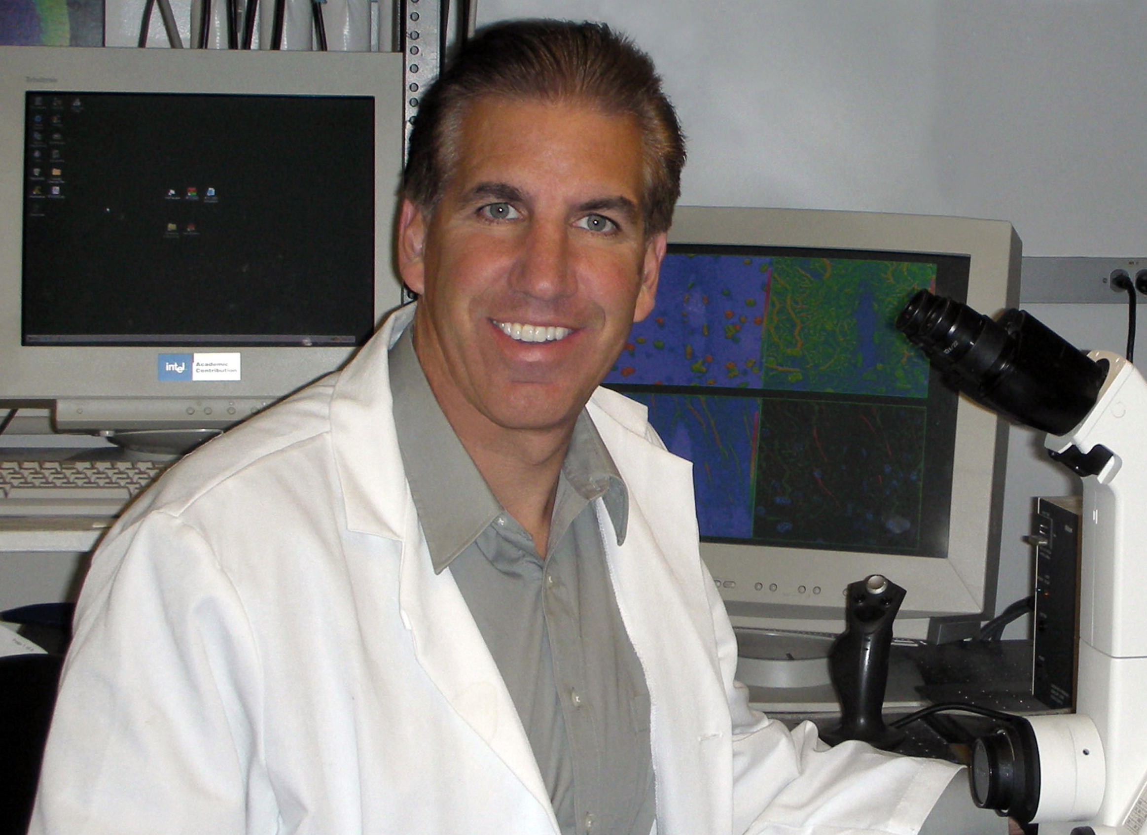
Photo courtesy Thomas Deerinck
Question: What’s your background?
Answer: "I was born and raised in Stockton from a family of 10 kids and I attended A.A. Stagg High School. When I was a senior, Dr. Betty Barbour (later Betty Mathews) came and gave a talk on the program she had recently started at Delta College that was one-of-a-kind: to train people to be electron microscopists. I was hooked almost immediately by the stunning scanning electron micrographs she showed."
Q: And what is the nature of your job today?
A: "After graduating I was recruited in 1978 to the Department of Neurosciences at UC San Diego, one of the top neuroscience departments in the world, where I have worked ever since. As a senior technical specialist, I worked as part of a team to advance all types of microscopy, whether they use light, electrons or even X-rays. Tremendous advances are being made to allow us to study how life works in health and disease in ways not previously possible.
"One of my current focuses these days as a technical consultant is on a relatively new technology that is being developed to map the structure of the brain at nanometer-scale resolution, with the ultimate goal of obtaining a wiring diagram of the human brain. This goal is still quite a way off but we are getting closer every day."
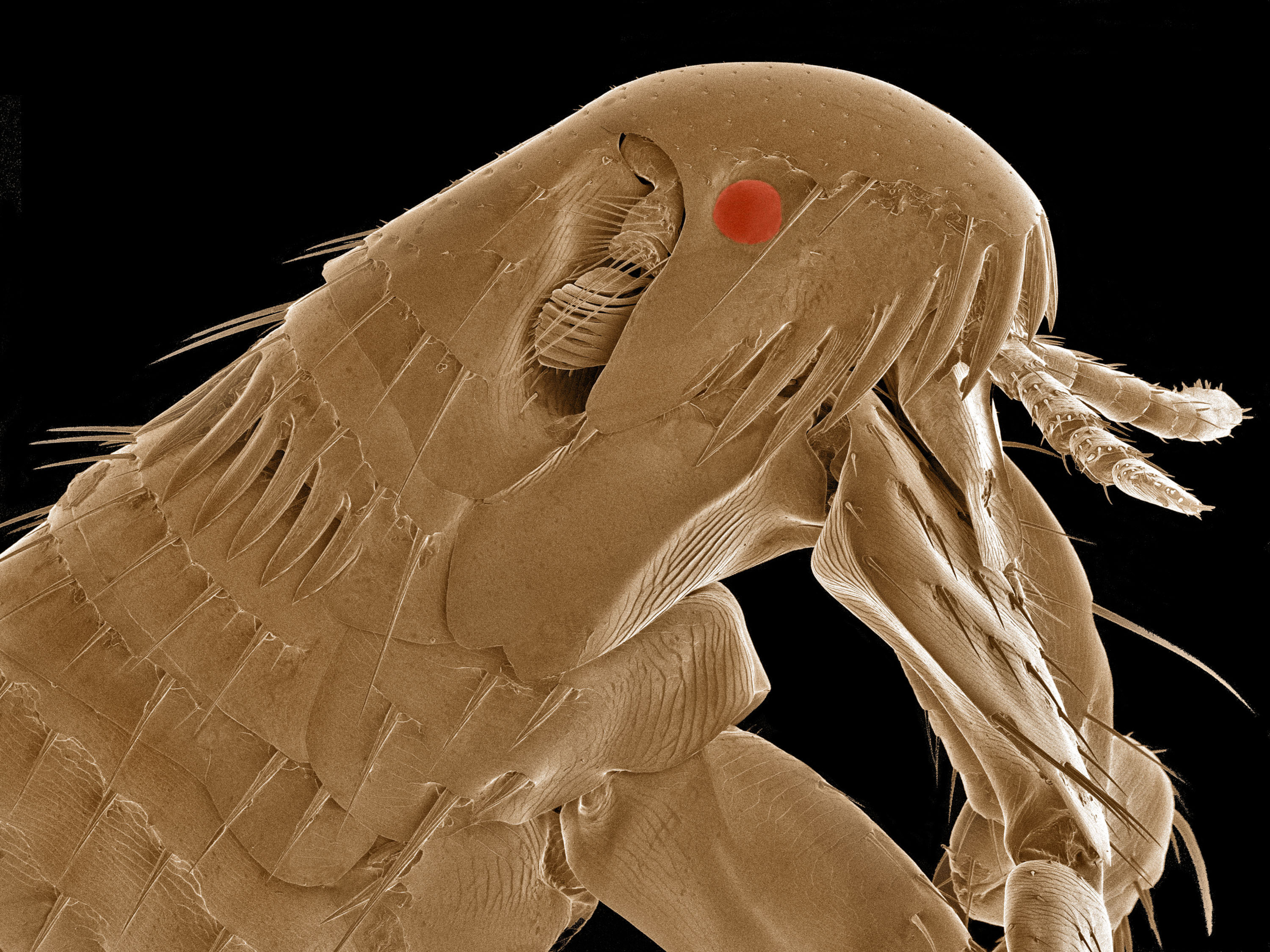
A common flea as seen by an electron microscope. Image created by Thomas Deerinck.
Q: What is it you enjoy about electron microscopy?
A: "Electron microscopes give you the power to image subjects with an almost unlimited magnification (up to over 1 million times!). It sort of transports you to an unexplored alien world of the microscopic. I have had the opportunity to publish research papers with a half dozen Nobel laureates, trying to extend the capabilities of what can be imaged using microscopes, with direct applications to aging, cancer, and Alzheimer’s disease, just to name a few. Microscopy-driven biomedical research is advancing our knowledge of diseases and potential treatments at an astonishing rate."
Q: Is this almost an art form for you?
A: "I am fortunate to be married for the last 30 years to an artist, my wife Karla, who graduated from UCLA. She has been a big inspiration in teaching me about color, content, form and aesthetics. The microscopic world is as beautiful as the natural world all around us. If you approach it as one does nature photography, the end result is more compelling."
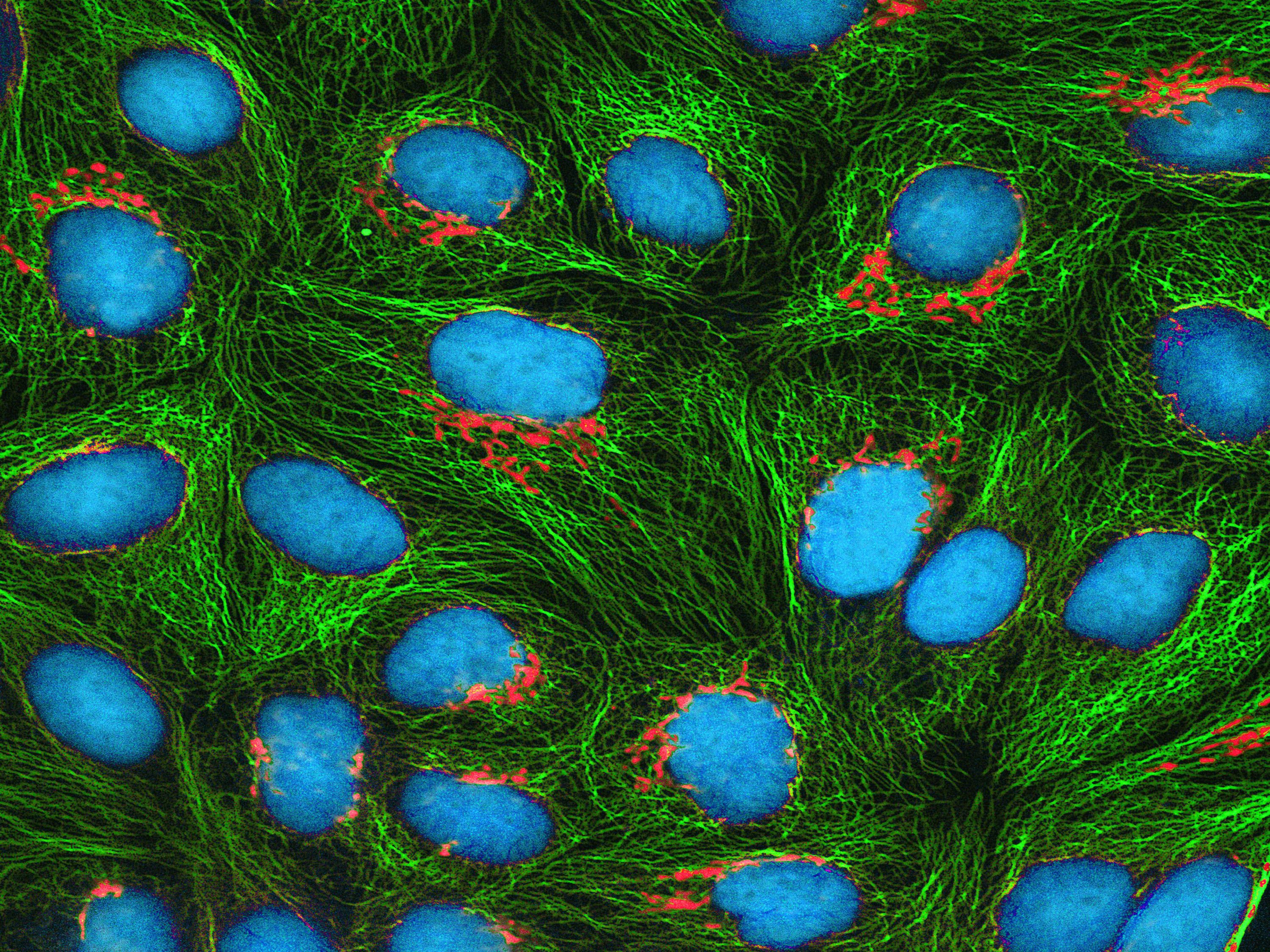
An image of HeLa cells, a human cell line used for research, as seen under an electron microscope. Image created by Thomas Deerinck.
Q: How unusual is it to have had this opportunity at the community college level?
A: "The program is extremely valuable in that graduates are in high demand in a wide range of disciplines, from university-level research labs, to pharmaceutical companies, to the semiconductor industry and even defense contractors. One can also very easily transfer after two years to a UC school to obtain a four-year degree at a fraction of the cost. Even without this, many Delta College-trained technicians move up quickly to head various research labs around the country."
Q: Any other thoughts on how Delta shaped your life’s direction?
A: "Coming from a family of 10, the program was a godsend. It allowed me to gain the training and expertise needed to enter the realm of biomedical research at a very high level."
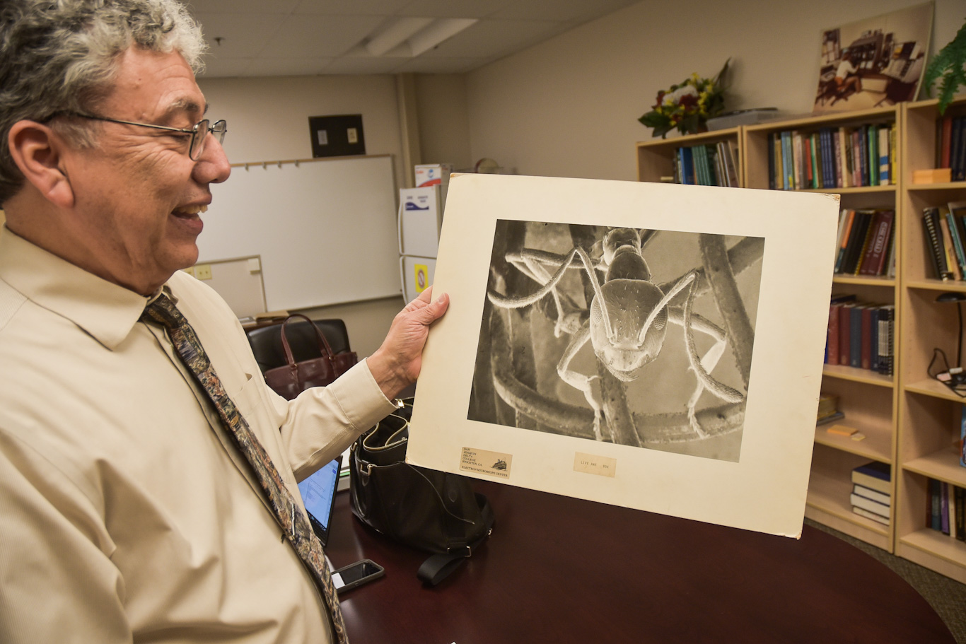
Electron microscopy Professor Frank Villalovoz holds an image of an ant that was created by Thomas Deerinck when he was a student at Delta more than 40 years ago.